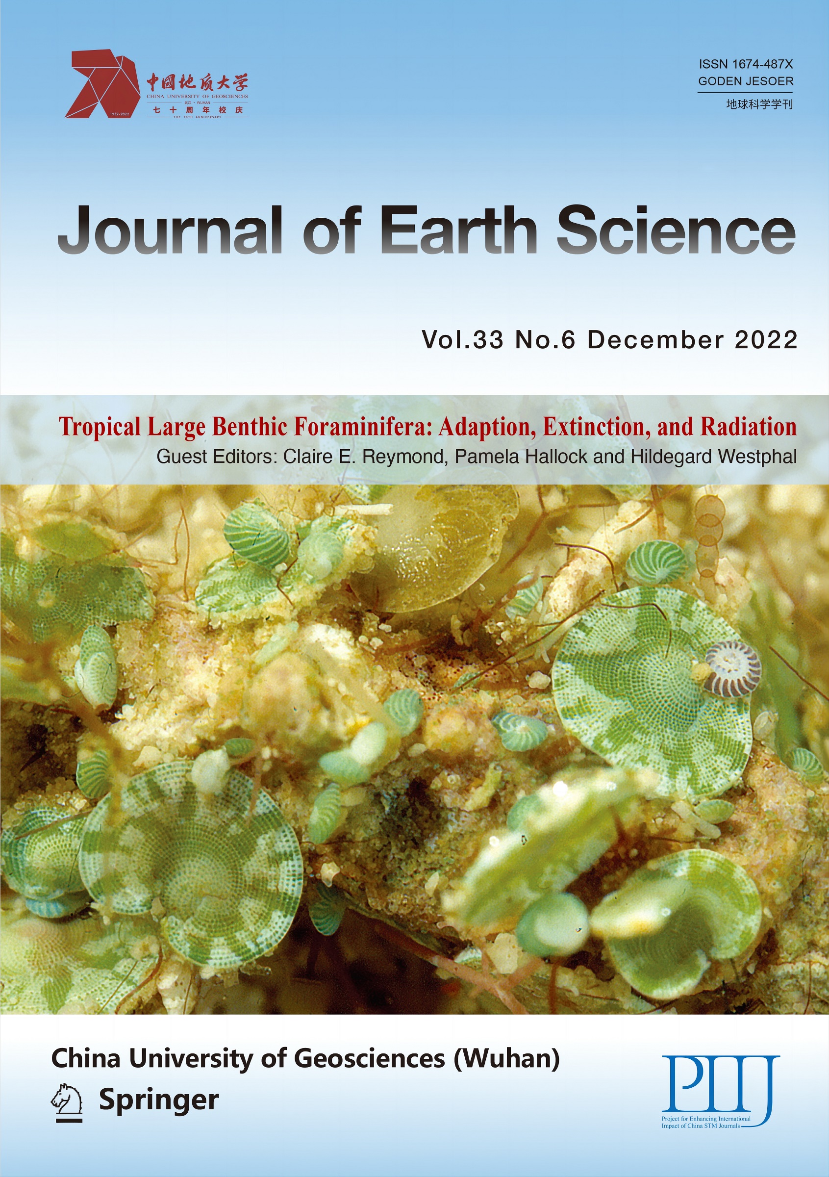
Foraminifera are highly diverse and have a long evolutionary history. As key bioindicators, their phylogenetic schemes are of great importance for paleogeographic applications, but may be hard to recognize correctly. The phylogenetic relationships within the prominent genus Amphistegina are still uncertain. Molecular studies on Amphistegina have so far only focused on genetic diversity within single species and suggested a cryptic diversity that demands for further investigations. Besides molecular sequencing-based approaches, different mass spectrometry-based proteomics approaches are increasingly used to give insights into the relationship between samples and organisms, especially as these do not require reference databases. To better understand the relationship of amphisteginids and test different proteomics-based approaches we applied de novo peptide sequencing and similarity clustering to several populations of Amphistegina lobifera, A. lessonii and A. gibbosa. We also analyzed the dominant photosymbiont community to study their influence on holobiont proteomes. Our analyses indicate that especially de novo peptide sequencing allows to reconstruct the relationship among foraminiferal holobionts, although the detected separation of A. gibbosa from A. lessonii and A. lobifera may be partly influenced by their different photosymbiont types. The resulting dendrograms reflect the separation in two lineages previously suggested and provide a basis for future studies.
The genus Amphistegina was first described by d'Orbigny (d'Orbigny, 1826) and then Ehrenberg (Ehrenberg, 1838). Current studies suggest that at least 11 extinct relatives (Larsen, 1978) have been widely distributed since the Eocene and throughout the Miocene (Todd, 1976), and it contains a minimum of six to seven commonly described recent morphospecies (Förderer and Langer, 2019; Renema, 2018; Debenay, 2013; Murray, 2006; Langer and Hottinger, 2000; Hottinger et al., 1993; Larsen, 1978). These are generally grouped in two morpho-groups, with lineage I (Larsen, 1978), or the A. radiata- group (Renema, 2018) containing the more symmetrical forms A. radiata and A. papillosa, as well as, according to Larsen (1978), also A. gibbosa (shown in Fig. 3) and A. bicirculata. Lineage II or the A. lessonii-group has widely spaced curved septa and is therefore more asymmertrical in axial view (Renema, 2018). It contains A. lessonii, A. lobifera (both shown in Fig. 3) and, according to e.g. Renema (2018), also A. bicirculata and A. quoi, of which the latter is rarely recognized by others. A. gibbosa is not described by Renema (2018), however, briefly mentioned as "western Atlantic sister species of A. lobifera and A. lessonii". Hohenegger (2014) distinguishes between the A. lessonii kinship and the A. radiata kinship like Renema (2018), though he does not mention the existence of A. gibbosa. Besides these, also A. madagascariensis is occasionally described (Förderer and Langer, 2019; Murray, 2006; Zmiri et al., 1974), but assumed to be a variety of A. lessonii (Förderer and Langer, 2019).
Most of these extant species are widely distributed throughout the Indo-Pacific and show a depth-related niche partitioning that is related to shape trends (Hohenegger, 2004; Hallock and Hansen, 1979), but partly overlaps, resulting in locally mixed assemblages (more details on species descriptions and distributions can be found in Förderer and Langer, 2019; Renema, 2018; Debenay, 2013; Murray, 2006; Loeblich and Tappan, 1994, 1988; Hottinger et al., 1993; Larsen, 1978; Hansen and Buchard, 1977). A. gibbosa, in contrast, is generally assumed to be the only Amphistegina species present in the Atlantic, specifically the Caribbean region. This is however controversial (Hallock, 1999) and some authors identified Amphistegina found along the coast of Brazil as A. lessonii instead (Alves and Júnior, 2020; Prazeres et al., 2012, 2011). In light of their ubiquitous appearance in shallow warm-water carbonate systems it is even more surprising that species boundaries and numbers remain widely uncertain (Renema, 2018) and that therefore a reassessment of their phylogenetic and evolutionary relationships is long overdue.
Molecular studies on Amphistegina have so far only focused on genetic diversity within single species. Schmidt et al. (2016) showed a distinction between A. lobifera from Israel (Mediterranean and Red Sea) and Australia (Great Barrier Reef). This was confirmed by a detailed study of Prazeres et al. (2020b), who differentiated between six genotypes of A. lobifera that could indeed represent at least four separate species and have distinct distribution ranges structured along a longitudinal gradient. Subsequently, Ramadhani et al. (2022, under review) determined three distinct genetic types of A. lessonii across its global range, with several of these co-occurring in the centre of its distribution. This further suggests a high cryptic diversity within the genus and demands for broader investigations of these prolific foraminifera using modern approaches.
Besides RNA and DNA sequencing-based molecular approaches, different mass spectrometry (MS)-based proteomics approaches are increasingly used to give insights into the relationship between samples and organisms (Aebersold and Mann, 2003). The most common method is to first generate short peptides and determine their mass-to-charge (m/z) ratios by MS. These measured mass spectra are then compared to a given database to find the matching peptide sequences and in turn identify the corresponding proteins. However, this method fails to recognize peptides that do not have existing sequence data in the given database, or for organisms that do not have a suitable reference database at all (MacCoss, 2005). This has limited the application of proteomics methods largely to the analysis of model organisms. However, in recent years instrumental and methodological advances enabled the adaption of these methods to a wider range of samples (Heck and Neely, 2020). In cases where reference databases are unavailable, de novo peptide sequencing, i.e., direct inference of peptide sequences from tandem mass spectra, is often the only option. Because this may have some limitations, a novel synergistic approach optimizes de novo peptide sequencing by combining the outcomes of several commonly available algorithms (Blank-Landeshammer et al., 2017). Another MS-based approach is to cluster similar peptide mass spectra by similarity, using different algorithms (Rieder et al., 2017b). These commonly used clustering approaches can be further improved by the DISMS2 filter, which allows to also consider precursor mass, precursor charge or retention time. By comparing thousands of peptide spectra, without de novo annotation or reference database, the similarities among sample proteomes are calculated and clustered, which may indicate their phylogenetic proximity (Rieder et al., 2017a).
To better understand the relationship of recent Amphistegina and at the same time test the different proteomics-based approaches on species with no appropriate reference database, we applied and compared the results of de novo peptide sequencing as well as clustering with DISMS2 to three recent Amphistegina species. These were collected from different sites, partly repeatedly over several years, to test for the influence of spatial and temporal variations. LBF are holobionts, i.e., a combination of the foraminiferal host organism, their photosymbiotic algae and microbiome. As these associated organisms were included in the proteomics workflow, they are also contained in the resulting MS data. We therefore applied a DNA sequencing approach to analyse the dominant photosymbiont community, and their potential influence on the holobiont similarity clusters.
1.
MATERIALS AND METHODS
1.1
Sample Collection
Samples were collected by snorkelling or SCUBA diving during different field campaigns (Stuhr et al., 2021, 2018b) following standard protocols in all locations (Red Sea (RS), Eilat, Israel; Western Indian Ocean (WIO), Zanzibar, Tanzania; Western Atlantic (WA), Florida Keys, USA), and either directly frozen at -80 ℃ or cultured at ZMT for several months (Table 1), in which case LBF were kept in sterilized seawater for three days prior to freezing in order to limit contamination by the microbiome within the culture dishes. They were then either extracted for DNA analysis in Bremen or transported on dry ice to ISAS, Dortmund, for proteomic analyses.
Table
1.
Site, depth, and year of collection of foraminifera populations analysed in this study
Species
Site
Year
Depth (m)
Collection
Abbreviation
Marginopora vertebralis
Zanzibar, WIO
2014
3
Culture
M. vertebralis WIO
Amphistegina lessonii
Eilat, RS
2016
3
Culture
A. lessonii RS16
Amphistegina lessonii
Eilat, RS
2016
3
Field
A. lessonii RS*16
Amphistegina lessonii
Zanzibar, WIO
2015
3
Culture
A. lessonii WIO15
Amphistegina lessonii
Zanzibar, WIO
2014
10
Culture
A. lessonii WIO14
Amphistegina lobifera
Eilat, RS
2016
3
Culture
A. lobifera RS16
Amphistegina lobifera
Eilat, RS
2016
3
Field
A. lobifera RS*16
Amphistegina lobifera
Zanzibar, WIO
2015
3
Culture
A. lobifera WIO15
Amphistegina gibbosa
Florida, WA
2015
18
Culture
A. gibbosa WA15
Amphistegina gibbosa
Florida, WA
2014
18
Culture
A. gibbosa WA15
Foraminifera frozen immediately after collection (field) are marked with an asterisk *.
Eight specimens per replicate were pooled and processed as described previously (Stuhr et al., 2018a; Rieder et al., 2017a). After protein digestion and quality-control, amino acid analysis was performed on all samples. Thus, sample amounts were corrected to compensate for systematic errors and to guarantee equal starting material for the subsequent LC-MS analysis.
1.2.2
LC-MS/MS analysis
Using an Ultimate 3000 nano-RSLC system coupled to an Orbitrap Fusion Lumos mass spectrometer (both Thermo Scientific), 330 ng per sample were analysed. A 100 µm × 2 cm C18 trapping column was used for preconcentration of peptides for 10 min using 0.1% TFA (v/v) with a flow rate of 20 µL/min followed by separation on a 75 µm × 50 cm C18 main column (both PepMap RSLC, Thermo Scientific) with a 90 min LC gradient ranging from 3%–35% of buffer B: 84% acetonitrile, 0.1% formic acid at a flow rate of 250 nL/min. The Orbitrap Fusion Lumos MS was operated in data-dependent acquisition mode and MS survey scans were acquired from m/z 300 to 1 500 at a resolution of 120 000 using the polysiloxane ion at m/z 445.12002 as lock mass. Isolation of precursors was performed by the quadrupole with a window of 0.4 m/z. Acquisition was performed in top speed mode, selecting the most intense signals within a cycle time of 3 s between survey scans and subjecting them to higher energy collisional dissociation with a normalized collision energy of 32% at a resolution of 15 000, taking into account a dynamic exclusion of 15 s. Automated gain control target values were set to 2 × 10⁵ for MS and 5 × 10⁴ MS/MS. Maximum injection times were 120 and 250 ms, respectively. Precursor ions with charge states of +1, > +7 or unassigned were excluded from MS/MS analysis and monoisoptic peak determination was set to 'peptide'.
1.2.3
Proteome-wide distance measure DISMS2
DISMS2 is a flexible algorithm that computes proteome-wide distances between MS/MS runs consisting of thousands of unidentified spectra, without additional information (Rieder et al., 2017a). Here, the ProteoWizard tool MSConvertGUI (Chambers et al., 2012) was used to convert Thermo RAW files into mzXML files. The open data format mzXML can be read with the R package readMzXmlData (Gibb, 2015). The washing step of the liquid chromatography was excluded. Thus, only spectra of the first hundred minutes are part of the analysis. First, distances for all these spectra were computed. Second, in each run 2 000 spectra with highest total ion signal were extracted. The preselection of spectra has already been used before (Palmblad and Deelder, 2012).
We used the same full factorial design as in the original paper (Rieder et al., 2017a) to find optimal parameter settings for all spectra. Out of in total 81 factor combinations the combination with the highest partial R squared (0.618 3) was chosen. Partial R squared is a goodness of fit measure that was calculated based on adonis, an ANOVA like approach for distance matrices. Optimized parameters correspond to the values from the original paper. In the pre-processing step no selection of highest peaks (topn = ∞) is made and binning is performed with binsize bin = 0.2. In the step that includes checking of constraints the size of retention time window is ret = 3 000 and the accepted precursor mass shift limit is prec = 10 ppm. The cosine distance (dist = dcos) is chosen as distance measure between spectra and the cutoff for distance of spectra is cdis = 0.3.
1.2.4
De novo peptide sequencing data analysis
Conversion of generated.raw files and subsequent data analysis by de novo peptide sequencing was performed as described by Blank-Landeshammer et al. (2017). Only full de novo sequences of agreeing spectrum annotations by PEAKS, pNovo+ and Novor—fulfilling an expected false discovery rate (FDR) of 5%—were used for further analysis.
For distance calculation, first the similarity of peptide identifications was determined by calculating the Sørensen-Dice coefficient (Sørensen, 1948; Dice, 1945) for all sample pairs, where |X| and |Y| represent the number of unique peptides in the samples.
QS=2|X⋂Y||X|+|Y|
For comparison of populations, all identified peptides within the replicates were pooled and compared as described above.
All statistical analyses were performed and figures were produced with the statistical programming software R (R Core Team, 2016), Version 4.1.0. Package readMzXmlData (Palmblad and Deelder, 2012) was used for reading MS data, and packages ape (Paradis et al., 2004) and phytools (Revell, 2012) were used for plotting dendrograms. In each case, dendrograms were generated by using complete linkage hierarchical clustering and triplicates of M. vertebralis WIO14 as an outgroup. The dendrograms of two different approaches were visualized facing each other for direct comparison.
1.3
Genetic Analyses
1.3.1
Molecular analysis of photosymbionts
Symbiont DNA was extracted and prepared for 18S sequencing as previously described in Stuhr et al. (2018b). Briefly, 36 foraminifera (n = 4 per population) were crushed with a micropestle and digested in a CTAP/protein kinase K lysis buffer, and subsequently extracted in phenol-chloroform (Green and Sambrook, 2012). After ethanol washing, a 443-bp fragment of the 3´end of the photosymbionts' 18S rDNA was amplified using primers SymFS1 and 1528R (Schmidt et al., 2015) and Taq polymerase using previously described cycling conditions (Stuhr et al., 2018b). PCR products were SAP digested and Sanger sequenced (Starseq, Mainz) without cloning. The sequences were trimmed for primers and with a quality threshold of 0.01 (1% error probability) in Geneious Prime 2021.1.1. The resulting contigs were deposited at ENA (project accession number PRJEB49701) and aligned after adding sequences with close BLAST hits or from diatoms previously described or sequenced from LBFs extracts (Prazeres et al., 2021; Stuhr et al., 2021, 2018b; Barnes, 2016; Lee, 2006). Alter (Glez-Peña et al., 2010) was used to condense haplotypes with identical sequences. MEGA X (Kumar et al., 2018) was used to determine the best fitting substitution model and finally the taxonomic assignment was done by calculating maximum- likelihood phylogenetic trees in RAxML (Kozlov et al., 2019).
2.
RESULTS
2.1
Proteome-Based Clustering
The comparison of dendrograms resulting from proteome-wide distance measures DISMS2 on either all or only the top 2 000 spectra (Fig. 1), illustrating distances between MS/MS runs of different LBF populations and their technical replicates, showed relatively similar overall results, grouping all samples according to species. However, if all spectra were included in the analysis (left side), the uncultured A. lessonii RS16 samples and one A. lessonii WIO15 sample grouped together with all A. lobifera, instead of with the remaining A. lessonii. In contrast, the A. lessonii WIO14 samples formed a separate cluster when using only the top 2 000 spectra for DISMS2 (right side), yet these did not merge with samples of other species. Samples also cluster more according to their collection site and year using this approach, e.g. with all A. lobifera triplicates grouping by their population and then by location. A. gibbosa formed a separate cluster in both approaches and hence clustered dominantly by population and then collection year. The average distance between technical replicates of the populations was between 40% and 60% when all spectra were considered and between 35% and 50% for the top 2 000 spectra. Nodes connecting all samples of each species were at ~80% and around 50%–70% distances, respectively.
Figure
1.
Proteome-wide distance measure clusters computed via DISMS2 with optimized parameters based on either pairwise distances of all spectra (left side) or only the top 2 000 spectra (right side) produced by 30 MS/MS runs of triplicates of different population of large benthic foraminifera, using complete linkage hierarchical clustering.
Since DISMS2 with the top 2 000 spectra showed a very coherent result regarding grouping of triplicates and populations, this was considered as the more reliable approach and further compared to the output of a de novo annotation approach. Complete linkage hierarchical clustering of de novo peptide sequences resulted in widely similar grouping (Fig. 2). One sample of A. lessonii WIO15 grouped more closely with A. lobifera than the other A. lessonii samples, which however clustered closer together without building a separate group for the population sampled in 2014. Moreover, within A. lobifera the samples of the cultured RS16 and WIO15 populations grouped first together, and then the uncultured RS16 samples. Apart from these minor differences, populations and triplicates within all LBF, including A. gibbosa and M. vertebralis, showed widely analogous clusters. In both approaches, A. lobifera and A. lessonii are clearly more similar to each other than they are to A. gibbosa. The average distance between technical replicates was between 35% and 50% for de novo peptide sequences. Nodes connecting all samples of each species were around 50%–60% distances.
Figure
2.
Proteome-wide distance measure clusters computed via de novo peptide sequences (left side) or DISMS2 on top 2 000 spectra with optimized parameters (right side) produced by 30 MS/MS runs of triplicates of different population of large benthic foraminifera, using complete linkage hierarchical clustering.
Because only one sample did not cluster with the remainder of its population and species, we regarded the de novo approach as providing the most representative results. When triplicates of each population were pooled, the resulting hierarchical cluster (Fig. 3) grouped robustly by LBF species, with A. lessonii and A. lobifera being most similar to each other, followed by A. gibbosa. Within species, no dominant grouping by collection site of year could be identified.
Figure
3.
Proteome-wide distance measure clusters computed via de novo peptide sequences produced by 30 MS/MS runs of pooled triplicates of different population of large benthic foraminifera, using complete linkage hierarchical clustering. Images on the right side show from top to bottom representative specimens of Amphistegina lessonii, A. lobifera and A. gibbosa. Marginopora vertebralis is not displayed.
To understand whether the similarity patterns of the tested LBF reconstructed by proteomics-based approaches may reflect their different photosymbiont communities, we applied a widely used DNA barcoding approach and calculated phylogenetic a tree of the resulting ~450 bp 18S rDNA sequences. This molecular fingerprinting of all samples (n = 24, Fig. 4) revealed that all A. gibbosa (n = 5) contained the same diatom symbiont sequence, while A. lessonii (n = 11) yielded seven haplotypes and A. lobifera (n = 8) yielded five haplotypes. All these sequences fall within the order Fragilariales that cluster closely together with sequences obtained from formerly sequenced A. lobifera from Israel, Indonesia, Australia, the Maldives and Papua New Guinea (Prazeres et al., 2021; Stuhr et al., 2021), A. lessonii from Zanzibar (WIO), Palau, Papua New Guinea, Australia and Eilat (RS)(Ramadhani et al., 2022, under review; Prazeres et al., 2021; Stuhr et al., 2018b) and A. gibbosa from Florida (WA) (Stuhr et al., 2018b). In contrast, A. radiata from Papua New Guinea (Prazeres et al., 2021) host endosymbionts from Triceratiales instead, another order of diatoms. Although the obtained sequences (443 bp) are too short for a taxonomic assignment to the species level, our results indicate some variability in the diatom assemblages, especially within A. lessonii. The resulting dendrogram also showed a separation of A. lobifera endosymbiont sequences from Greece and A. gibbosa photosymbionts from the other samples, while A. lessonii and A. lobifera photosymbionts grouped weakly by collection sites. The minor division of greek A. lobifera from the remaining samples could be due to a change in their main associated pennate diatoms during the spreading of the species on its northwestward expansion in the Mediterranean (Guastella et al., 2019; Langer et al., 2013; Triantaphyllou et al., 2009) as an adjustment to varying environmental conditions or native diatom communities. While Schmidt et al. (2016) found no systematic difference in the photosymbiont community of Red Sea and Mediterranean populations, the diatom population in other areas, potentially more marginal in terms of A. lobifera's temperature range, are likely to bear a different local diatom community with which the foraminifera can associate. Moreover, it is not clear whether all A. lobifera in the Mediterranean are really Lessepsian invaders or might have actually been present in lower densities already before the opening of the Suez canal (Langer, 2008), in which case they could have adjusted their photosymbiont community to the different environmental conditions over much longer time scales.
Figure
4.
Phylogenetic assignment of diatom-symbiont sequences from Amphistegina lobifera and A. lessonii collected in Zanzibar, Tanzania, and Eilat, Israel, or A. gibbosa from the Florida Keys, USA. These new sequences are highlighted in bold and underlined. Bootstrap support above 50% is given at the respective nodes.
The results of both our proteomics-based approaches showed a separation of the three species. However, as DISMS2 applied to all spectra inadequately grouped the uncultured A. lessonii RS16 samples and one A. lessonii WIO15 sample with A. lobifera, this approach gives the least reliable results. This is further indicated by the highest distances between population replicates as well as nodes connecting all samples of the same species (excluding described faulty grouped samples).
Although these distances were lower when using the top 2 000 spectra only, the A. lessonii WIO14 formed a group separated from the other A. lessonii and also the A. lobifera samples. Hence, the de novo peptide sequences-based dendrogram represented the most trustworthy outcome, with all but one sample (A. lessonii WIO15.2) clustering correctly by species, and principally also the technical triplicates by population, with comparably low distances. This might be because the de novo peptide sequencing algorithms act as efficient filters to separate true (i.e., biologically relevant) spectra from background spectra and those stemming from laboratory contaminations compared to unsupervised clustering. We therefore demonstrate that the approach of Blank-Landeshammer et al. (2017) is capable of comprehensively delineating LBF taxa without a reference database. Yet, as multiple algorithms can be easily applied to the same MS/MS dataset, a combination with DISMS2 (Rieder et al., 2017a) applied to top spectra may serve to further confirm the outcome and boost reliability.
The resultant dendrograms provide first hints into the phylogeny of the three studied Amphistegina species. Clearly, the Indopacific species A. lobifera and A. lessonii are more closely related to each other than they are to their counterpart A. gibbosa in the Atlantic (Figs. 1 to 3). This confirms that the morphologically relatively similar and therefore sometimes confused species A. lessonii and A. gibbosa are indeed separate species, and agrees with their phylogenetic separation suggested by Larsen (1978) into relatively widely separated lineages. This pattern is reflected in the photosymbiotic diatoms harbored by these populations (Fig. 4). Hence, the differences detected for the LBF holobionts may be influenced and partly based on the differences of the photosymbiont communities. Overall, as previously recognized, A. lessonii and A. lobifera contain a more diverse community of Fragilariales (Ramadhani et al., 2022, under review; Prazeres et al., 2021; Stuhr et al., 2021, 2018b; Barnes, 2016; Schmidt et al., 2016), while A. gibbosa contains only one strain of a Fragilariales species (Stuhr et al., 2018b; Barnes, 2016), fairly distinct from those harboured by the aforementioned species. Interestingly, also A. radiata appears to have a higher specificity in its photosymbiont assemblages, which is a member of Triceratiales instead of Fragilariales (Prazeres et al., 2021; Barnes, 2016). Whiles this may be an adaptation to their usually more light-reduced habitat (Prazeres et al., 2021), the variation in symbiont flexibility could likewise reflect their phylogenetic distinction into the two lineages suggested by Larsen (1978), with more restricted associations in lineage I (A. radiata-A. gibbosa) and highly flexible associations in lineage II (A. lessonii-A. lobifera). In this regard, it would be highly interesting to examine the photosymbiont communities of A. papillosa, which is generally assumed to belong to lineage I or the A. radiata group/kinship, to see whether it also shows a relatively high symbiont specificity.
Despite a potential effect of the global symbiont community on the proteome-based similarity clusters, the well-defined separation of morpho-species signifies the LBF host as the major driver. If the symbionts had an overriding impact, A. lobifera and A. lessonii as well as samples with morpho-species should cluster more mixed, as specimens from different collection sites or years showed similar symbiont sequences. In contrast, recent environmental conditions appear to have a detectable influence. For example, A. lobifera and A. lessonii from the Red Sea that were cultured prior to sampling (RS16) are more similar to their likewise cultured counterparts from the western Indian Ocean (WIO15) than they are to subsamples of the same Red Sea population sampled immediately without culturing (RS16*). Since the proteome is largely influenced by environmental factors and may thereby influence evolutionary processes itself (Baer and Millar, 2016) it appears reasonable that populations cultivated under alike conditions will have more similar proteomes. Conversely, A. lessonii WIO14 was in all dendrograms slightly disjointed from the remaining A. lessonii populations. Notably, it is the only A. lessonii population that was sampled in at least 10 m depth, while all others originated from 2–3 m depth. Whether this distinction in A. lessonii from Zanzibar may thus represent a genotypic differentiation related to their original habitat, or if this population was somehow differently affected by culturing, will have to be clarified in future studies. Conditions such as the microbiome or phenological phases can potentially vary between cultures, even under similar light, temperature, and water geochemistry, especially if these have been developing over long time.
4.
CONCLUSIONS
Our proteome-based clustering approaches along with phylogenetic analyses of their photosymbiont communities indicate that especially de novo peptide sequencing provides a reliable tool to reconstruct the evolutionary relationship among foraminiferal holobionts. Although the wide separation of A. gibbosa from A. lessonii and A. lobifera along with their distinctive type of Fragilariales symbionts may indicate a partial influence of the photosymbiont communities, the well-defined species separation in between A. lobifera and A. lessonii contradicts their highly widely overlapping symbiont communities. This difference between proteome-based clusters and the phylogenetic dendrogram of the photosymbionts therefore signifies that the symbiont community cannot be the major clustering driver. This further confirms that proteomics approaches allow us to draw phylogenetic information, which here reproduces the separation in two lineages as suggested by Larsen (1978). This study provides a basis for future studies that should include the remaining Amphistegina morpho-species and include populations from their wide spatial distribution, across depth and their entire geographic range, but should moreover also be extended to other genera of foraminifera. Shedding light onto the true (cryptic) diversity and unravelling the evolutionary history of the prominent genus Amphistegina will help ecologists and palaeontologists alike towards understanding paradigms in the face of changing environmental conditions, past and future adaptions, or potential radiation events.
ACKNOWLEDGMENTS:
This project was funded by the Leibniz Association (No. SAW-2014-ISAS-2) awarded to Hildegard Westphal, Albert Sickmann and Jörg Rahnenführer. Sampling in the Florida Keys was conducted under the Research Permit (No. FKNMS-2015-026). Samples from Zanzibar were collected under the Revolutionary Government of Zanzibar 434 (S.M.Z.) Research Permit (No. 0671). We sincerely thank Pamela Hallock and Gita R. Narayan for their sampling support, and the staff of the MAREE at ZMT for help with foraminifera cultures. The final publication is available at Springer via https://doi.org/10.1007/s12583-022-1609-1.
Alves, B. M., Júnior, M. N., 2020. Foraminiferal Assemblage Structure from Brazilian Tropical Urbanized Beaches (~7°S). Anais da Academia Brasileira de Ciências, 92(2): 1–15. https://doi.org/10.1590/0001-3765202020190486
Barnes, K. H., 2016. Diversity and Distribution of Diatom Endosymbionts in Amphistegina spp. (Foraminifera) Based on Molecular and Morphological Techniques: [Dissertation]. University of South Florida, St. Petersburg, Florida. 160
Bentov, S., Brownlee, C., Erez, J., 2009. The Role of Seawater Endocytosis in the Biomineralization Process in Calcareous Foraminifera. Proceedings of the National Academy of Sciences of the United States of America, 106(51): 21500–21504. https://doi.org/10.1073/pnas. 0906636106 doi: 10.1073/pnas.0906636106
Bhatt, K. A., Trivedi, M. H., 2018. Molecular Studies on Foraminifers: Past, Present, and Future. Journal of Foraminiferal Research, 48(3): 193–209. https://doi.org/10.2113/gsjfr.48.3.193
Blank-Landeshammer, B., Kollipara, L., Biß, K., et al., 2017. Combining de Novo Peptide Sequencing Algorithms, a Synergistic Approach to Boost both Identifications and Confidence in Bottom-up Proteomics. Journal of Proteome Research, 16(9): 3209–3218. https://doi.org/10.1021/acs.jproteome.7b00198
Boudagher-Fadel, M. K., 2018. Evolution and Geological Significance of Larger Benthic Foraminifera. Second Edition. UCL Press, London. 693. https://doi.org/10.2307/j.ctvqhsq3
Carnahan, E. A., Hoare, A. M., Hallock, P., et al., 2009. Foraminiferal Assemblages in Biscayne Bay, Florida, USA: Responses to Urban and Agricultural Influence in a Subtropical Estuary. Marine Pollution Bulletin, 59(8–12): 221–233. https://doi.org/10.1016/j.marpolbul.2009.08.008
Chambers, M. C., Maclean, B., Burke, R., et al., 2012. A Cross-Platform Toolkit for Mass Spectrometry and Proteomics. Nature Biotechnology, 30(10): 918–920. https://doi.org/10.1038/nbt.2377
d'Orbigny, A. D., 1826. Tableau Méthodique de la Classe des Céphalopodes. Annales des Sciences Naturelles, 7: 96–314
Debenay, J. -P., 2013. A Guide to 1 000 Foraminifera from Southwestern Pacific, New Caledonia. Muséum National d'Histoire Naturelle, Paris. 384
Dice, L., 1945. Measures of the Amount of Ecologic Association between Species. Ecology, 26(3): 297–302. https://doi.org/10.2307/1932409
Doo, S. S., Hamylton, S., Byrne, M., 2012. Reef-Scale Assessment of Intertidal Large Benthic Foraminifera Populations on one Tree Island, Great Barrier Reef and Their Future Carbonate Production Potential in a Warming Ocean. Zoological Studies, 51(8): 1298–1307. https://doi.org/10.1007/s00435-012-0164-1
Ehrenberg, C. G., 1838. Über die Bildung der Kreidefelsen und des Kreidemergels Durch Unsichtbare Organismen. Abhandlungen der Königlichen Akademie der Wissenschaften zu Berlin, Berlin. 59–147
Emrich, K., Martinez-Colon, M., Alegria, H., 2017. Is Untreated Sewage Impacting Coral Reefs of Caye Caulker, Belize? The Journal of Foraminiferal Research, 47(1): 20–33. https://doi.org/10.2113/gsjfr.47.1.20
Flakowski, J., Bolivar, I., Fahrni, J., et al., 2005. Actin Phylogeny of Foraminifera. Journal of Foraminiferal Research, 35(2): 93–102. https://doi.org/10.2113/35.2.93
Förderer, M., Langer, M. R., 2019. Exceptionally Species-Rich Assemblages of Modern Larger Benthic Foraminifera from Nearshore Reefs in Northern Palawan (Philippines). Revue de Micropaléontologie, 65: 100387. https://doi.org/10.1016/j.revmic.2019.100387
Förderer, M., Rödder, D., Langer, M. R., 2018. Patterns of Species Richness and the Center of Diversity in Modern Indo-Pacific Larger Foraminifera. Scientific Reports, 8(1): 8189. https://doi.org/10.1038/s41598-018-26598-9
Glas, M. S., Fabricius, K. E., de Beer, D., et al., 2012. The O2, pH and Ca2+ Microenvironment of Benthic Foraminifera in a High CO2 World. PLoS One, 7(11): e50010. https://doi.org/10.1371/journal.pone.0050010
Glez-Peña, D., Gómez-Blanco, D., Reboiro-Jato, M., et al., 2010. ALTER: Program-Oriented Conversion of DNA and Protein Alignments. Nucleic Acids Research, 38: W14–W18. https://doi.org/10.1093/nar/gkq321
Green, M. R., Sambrook, J., 2012. Molecular Cloning: A Laboratory Manual. Cold Spring Harbor Laboratory Press, Cold Spring Harbor, N. Y. 2028
Guastella, R., Marchini, A., Caruso, A., et al., 2019. "Hidden Invaders" Conquer the Sicily Channel and Knock on the Door of the Western Mediterranean Sea. Estuarine, Coastal and Shelf Science, 225: 106234. https://doi.org/10.1016/j.ecss.2019.05.016
Hallock, P., 1999. Symbiont-Bearing Foraminifera. In: Sen Gupta, B. K., ed., Modern Foraminifera. Kluwer Academic Publishers. 123–368
Hallock, P., Forward, L. B., Hansen, H. J., 1986. Influence of Environment on the Test Shape of Amphistegina. The Journal of Foraminiferal Research, 16(3): 224–231. https://doi.org/10.2113/gsjfr.16.3.224
Hallock, P., Glenn, E. C., 1986. Larger Foraminifera: A Tool for Paleoenvironmental Analysis of Cenozoic Carbonate Depositional Facies. PALAIOS, 1(1): 55–64. https://doi.org/10.2307/3514459
Hallock, P., Hansen, J., 1979. Depth Adaptation in Amphistegina: Change in Lamellar Thickness. Bulletin of the Geological Society of Denmark, 27: 99–104. https://doi.org/10.37570/bgsd-1978-27-10
Hallock, P., Lidz, B. H., Cockey-Burkhard, E. M., et al., 2003. Foraminifera as Bioindicators in Coral Reef Assessment and Monitoring: The Foram Index. Environmental Monitoring and Assessment, 81(1/2/3): 221–238. https://doi.org/10.1023/a:1021337310386
Hallock, P., Talge, H. K., Smith, K., et al., 1993. Bleaching in a Reef-Dwelling Foraminifer, Amphistengina Gibbosa. Proceedings of the 7th International Coral Reef Symposium, 1: 44–49
Hansen, H. J., Buchard, B., 1977. Depth Distribution of Amphistegina in the Gulf of Elat, Israel. Utrecht Micropaleontological Bulletins, 1977: 205–224
Haynes, J. R., 1992. Supposed Pronounced Ecophenotypy in Foraminifera. Journal of Micropalaeontology, 11(1): 59–63. https://doi.org/10.1144/jm.11.1.59
Heck, M., Neely, B. A., 2020. Proteomics in Non-Model Organisms: A New Analytical Frontier. Journal of Proteome Research, 19(9): 3595–3606. https://doi.org/10.1021/acs.jproteome.0c00448
Hohenegger, J., 2014. Species as the Basic Units in Evolution and Biodiversity: Recognition of Species in the Recent and Geological Past as Exemplified by Larger Foraminifera. Gondwana Research, 25(2): 707–728. https://doi.org/10.1016/j.gr.2013.09.009
Hohenegger, J., 2004. Depth Coenoclines and Environmental Considerations of Western Pacific Larger Foraminifera. The Journal of Foraminiferal Research, 34(1): 9–33. https://doi.org/10.2113/0340009
Hottinger, L., Halicz, E., Reiss, Z., 1993. Recent Foraminiferida from the Gulf of Aqaba, Red Sea. Slovenian Academy of Sciences and Arts and Swiss Academy of Natural Sciences, Ljubljana. 181
Kozlov, A. M., Darriba, D., Flouri, T., et al., 2019. RAxML-NG: A Fast, Scalable and User-Friendly Tool for Maximum Likelihood Phylogenetic Inference. Bioinformatics, 35(21): 4453–4455. https://doi.org/10.1093/bioinformatics/btz305
Kumar, S., Stecher, G., Li, M., et al., 2018. MEGA X: Molecular Evolutionary Genetics Analysis across Computing Platforms. Molecular Biology and Evolution, 35(6): 1547–1549. https://doi.org/10.1093/molbev/msy096
Langer, M., 2008. Foraminifera from the Mediterranean and the Red Sea. In: Aqaba-Eilat, the Improbable Gulf: Environment, Biodiversity and Preservation. Magnes Press, Jerusalem. 397–415
Langer, M. R., Hottinger, L., 2000. Biogeography of Selected "Larger" Foraminifera. Micropaleontology, 46: 105–126
Langer, M. R., Silk, M. T., Lipps, J. H., 1997. Global Ocean Carbonate and Carbon Dioxide Production: The Role of Reef Foraminifera. The Journal of Foraminiferal Research, 27(4): 271–277. https://doi.org/10.2113/gsjfr.27.4.271
Langer, M. R., Weinmann, A. E., Lötters, S., et al., 2013. Climate-Driven Range Extension of Amphistegina (Protista, Foraminiferida): Models of Current and Predicted Future Ranges. PLoS One, 8(2): e54443. https://doi.org/10.1371/journal.pone.0054443
Larsen, A. R., 1978. Phylogenetic and Paleobiogeographical Trends in the Foraminiferal genus Amphistegina. Revista Española de Micropaleontología, X: 217–243
Macher, J. N., Prazeres, M., Taudien, S., et al., 2021. Integrating Morphology and Metagenomics to Understand Taxonomic Variability of Amphisorus (Foraminifera, Miliolida) from Western Australia and Indonesia. PLoS One, 16(1): e0244616. https://doi.org/10.1371/journal.pone.0244616
Morard, R., Füllberg, A., Brummer, G. J. A., et al., 2019. Genetic and Morphological Divergence in the Warm-Water Planktonic Foraminifera Genus Globigerinoides. PLoS One, 14(12): e0225246. https://doi.org/10.1371/journal.pone.0225246
Mouanga, G. H., Langer, M. R., 2014. At the Front of Expanding Ranges: Shifting Community Structures at Amphisteginid Species Range Margins in the Mediterranean Sea. Neues Jahrbuch Für Geologie Und Paläontologie-Abhandlungen, 271(2): 141–150. https://doi.org/10.1127/0077-7749/2014/0381
Murray, J., 2006. Ecology and Applications of Benthic Foraminifera. Palaeogeography, Palaeoclimatology, Palaeoecology, 95: 1–426. https://doi.org/10.1017/cbo9780511535529
Narayan, G. R., Reymond, C. E., Stuhr, M., et al., 2022. Response of Large Benthic Foraminifera to Climate and Local Changes: Implications for Future Carbonate Production. Sedimentology, 69(1): 121–161. https://doi.org/10.1111/sed.12858
Narayan, Y. R., Lybolt, M., Zhao, J. X., et al., 2015. Holocene Benthic Foraminiferal Assemblages Indicate Long-Term Marginality of Reef Habitats from Moreton Bay, Australia. Palaeogeography, Palaeoclimatology, Palaeoecology, 420: 49–64. https://doi.org/10.1016/j.palaeo.2014.12.010
Palmblad, M., Deelder, A. M., 2012. Molecular Phylogenetics by Direct Comparison of Tandem Mass Spectra. Rapid Communications in Mass Spectrometry, 26(7): 728–732. https://doi.org/10.1002/rcm.6162
Paradis, E., Claude, J., Strimmer, K., 2004. APE: Analyses of Phylogenetics and Evolution in R Language. Bioinformatics, 20(2): 289–290. https://doi.org/10.1093/bioinformatics/btg412
Pawlowski, J., Holzmann, M., 2008. Diversity and Geographic Distribution of Benthic Foraminifera: A Molecular Perspective. Biodiversity and Conservation, 17(2): 317–328. https://doi.org/10.1007/s10531-007-9253-8
Pawlowski, J., Holzmann, M., 2002. Molecular Phylogeny of Foraminifera a Review. European Journal of Protistology, 38(1): 1–10. https://doi.org/10.1078/0932-4739-00857
Pawlowski, J., Holzmann, M., Tyszka, J., 2013. New Supraordinal Classification of Foraminifera: Molecules Meet Morphology. Marine Micropaleontology, 100: 1–10. https://doi.org/10.1016/j.marmicro.2013. 04.002 doi: 10.1016/j.marmicro.2013.04.002
Pawlowski, J., Lecroq, B., 2010. Short rDNA Barcodes for Species Identification in Foraminifera. Journal of Eukaryotic Microbiology, 57(2): 197–205. https://doi.org/10.1111/j.1550-7408.2009.00468.x
Pawlowski, J., Lejzerowicz, F., Esling, P., 2014. Next-Generation Environmental Diversity Surveys of Foraminifera: Preparing the Future. The Biological Bulletin, 227(2): 93–106. https://doi.org/10.1086/bblv227n2p93
Pisapia, C., Kateb, A. E., Hallock, P., et al., 2017. Assessing Coral Reef Health in the North Ari Atoll (Maldives) Using the FoRAM Index. Marine Micropaleontology, 133: 50–57. https://doi.org/10.1016/j.marmicro.2017.06.001
Pochon, X., Montoya-Burgos, J. I., Stadelmann, B., et al., 2006. Molecular Phylogeny, Evolutionary Rates, and Divergence Timing of the Symbiotic Dinoflagellate Genus Symbiodinium. Molecular Phylogenetics and Evolution, 38(1): 20–30. https://doi.org/10.1016/j.ympev.2005.04.028
Prazeres, M., Martínez-Colón, M., Hallock, P., 2020a. Foraminifera as Bioindicators of Water Quality: The FoRAM Index Revisited. Environmental Pollution, 257: 113612. https://doi.org/10.1016/j.envpol.2019.113612
Prazeres, M., Morard, R., Roberts, T. E., et al., 2020b. High Dispersal Capacity and Biogeographic Breaks Shape the Genetic Diversity of a Globally Distributed Reef-Dwelling Calcifier. Ecology and Evolution, 10(12): 5976–5989. https://doi.org/10.1002/ece3.6335
Prazeres, M., Renema, W., 2019. Evolutionary Significance of the Microbial Assemblages of Large Benthic Foraminifera. Biological Reviews, 94(3): 828–848. https://doi.org/10.1111/brv.12482
Prazeres, M., Roberts, T. E., Ramadhani, S. F., et al., 2021. Diversity and Flexibility of Algal Symbiont Community in Globally Distributed Larger Benthic Foraminifera of the Genus Amphistegina. BMC Microbiology, 21(1): 243. https://doi.org/10.1186/s12866-021-02299-8
Prazeres, M., Martins, S. E., Bianchini, A., 2011. Biomarkers Response to Zinc Exposure in the Symbiont-Bearing Foraminifer Amphistegina Lessonii (Amphisteginidae, Foraminifera). Journal of Experimental Marine Biology and Ecology, 407(1): 116–121. https://doi.org/10.1016/j.jembe.2011.07.015
Prazeres, M., Martins, S. E., Bianchini, A., 2012. Assessment of Water Quality in Coastal Waters of Fernando de Noronha, Brazil: Biomarker Analyses in Amphistegina Lessonii. The Journal of Foraminiferal Research, 42(1): 56–65. https://doi.org/10.2113/gsjfr.42.1.56
Prazeres, M., Uthicke, S., Pandolfi, J. M., 2015. Ocean Acidification Induces Biochemical and Morphological Changes in the Calcification Process of Large Benthic Foraminifera. ProceedingsBiological Sciences, 282: 8–11. https://doi.org/10.1098/rspb.2014.2782
Ramadhani, S. F., Roberts, T. E., Stuhr, M., et al., 2022. Patterns of Host-Symbiont Genetic Diversity and Specificity in the Reef-Dwelling Large Benthic Foraminifera Amphistegina lessonii. Coral Reefs, Under Review
Renema, W., 2003. Larger Foraminifera on Reefs around Bali (Indonesia). Zool Verh Leiden, 345: 337–366
Renema, W., 2018. Terrestrial Influence as a Key Driver of Spatial Variability in Large Benthic Foraminiferal Assemblage Composition in the Central Indo-Pacific. Earth-Science Reviews, 177: 514–544. https://doi.org/10.1016/j.earscirev.2017.12.013
Revell, L. J., 2012. Phytools: An R Package for Phylogenetic Comparative Biology (and other Things). Methods in Ecology and Evolution, 3(2): 217–223. https://doi.org/10.1111/j.2041-210X.2011.00169.x
Rieder, V., Blank-Landeshammer, B., Stuhr, M., et al., 2017a. DISMS2: A Flexible Algorithm for Direct Proteome-Wide Distance Calculation of LC-MS/MS Runs. BMC Bioinformatics, 18(1): 148. https://doi.org/10.1186/s12859-017-1514-2
Rieder, V., Schork, K. U., Kerschke, L., et al., 2017b. Comparison and Evaluation of Clustering Algorithms for Tandem Mass Spectra. Journal of Proteome Research, 16(11): 4035–4044. https://doi.org/10.1021/acs.jproteome.7b00427
Ross, B. J., Hallock, P., 2014. Chemical Toxicity on Coral Reefs: Bioassay Protocols Utilizing Benthic Foraminifers. Journal of Experimental Marine Biology and Ecology, 457: 226–235. https://doi.org/10.1016/j.jembe.2014.04.020
Ross, B. J., Hallock, P., 2018. Challenges in Using Cell Tracker Green on Foraminifers that Host Algal Endosymbionts. PeerJ, 6: e5304. https://doi.org/10.7717/peerj.5304
Schmidt, C., Heinz, P., Kucera, M., et al., 2011. Temperature-Induced Stress Leads to Bleaching in Larger Benthic Foraminifera Hosting Endosymbiotic Diatoms. Limnology and Oceanography, 56(5): 1587–1602. https://doi.org/10.4319/lo.2011.56.5.1587
Schmidt, C., Morard, R., Almogi-Labin, A., et al., 2015. Recent Invasion of the Symbiont-Bearing Foraminifera Pararotalia into the Eastern Mediterranean Facilitated by the Ongoing Warming Trend. PLoS One, 10(8): e0132917. https://doi.org/10.1371/journal.pone.0132917
Schmidt, C., Morard, R., Prazeres, M., et al., 2016. Retention of High Thermal Tolerance in the Invasive Foraminifera Amphistegina Lobifera from the Eastern Mediterranean and the Gulf of Aqaba. Marine Biology, 163(11): 1–13. https://doi.org/10.1007/s00227-016-2998-4
Sørensen, T., 1948. Method of Establishing Groups of Equal Amplitude in Plant Sociology Based on Similarity of Species Content and Its Application to Analyses of the Vegetation on Danish Commons. Kongelige Danske Videnskabernes Selskab, Biologiske Skrifter, 5: 1–34
Stainbank, S., Spezzaferri, S., Beccari, V., et al., 2020. Photic Stress on Coral Reefs in the Maldives: The Amphistegina Bleaching Index. Ecological Indicators, 113: 106257. https://doi.org/10.1016/j.ecolind.2020.106257
Stuhr, M., Blank-Landeshammer, B., Reymond, C. E., et al., 2018a. Disentangling Thermal Stress Responses in a Reef-Calcifier and Its Photosymbionts by Shotgun Proteomics. Scientific Reports, 8: 3524. https://doi.org/10.1038/s41598-018-21875-z
Stuhr, M., Meyer, A., Reymond, C. E., et al., 2018b. Variable Thermal Stress Tolerance of the Reef-Associated Symbiont-Bearing Foraminifera Amphistegina Linked to Differences in Symbiont Type. Coral Reefs, 37(3): 811–824. https://doi.org/10.1007/s00338-018-1707-9
Stuhr, M., Cameron, L. P., Blank-Landeshammer, B., et al., 2021. Divergent Proteomic Responses Offer Insights into Resistant Physiological Responses of a Reef-Foraminifera to Climate Change Scenarios. Oceans, 2(2): 281–314. https://doi.org/10.3390/oceans2020017
Stuhr, M., Reymond, C. E., Rieder, V., et al., 2017. Reef Calcifiers are Adapted to Episodic Heat Stress but Vulnerable to Sustained Warming. PLoS One, 12(7): e0179753.https://doi.org/10.1371/journal.pone. 0179753 doi: 10.1371/journal.pone.0179753
Stulpinaite, R., Hyams-Kaphzan, O., Langer, M. R., 2020. Alien and Cryptogenic Foraminifera in the Mediterranean Sea: A Revision of Taxa as Part of the EU 2020 Marine Strategy Framework Directive. Mediterranean Marine Science, 21: 719–758. https://doi.org/10.12681/mms.24673
Talge, H. K., Hallock, P., 1995. Cytological Examination of Symbiont Loss in a Benthic Foraminifera, Amphistegina Gibbosa. Marine Micropaleontology, 26(1/2/3/4): 107–113. https://doi.org/10.1016/0377-8398(95)00015-1
ter Kuile, B., Erez, J., 1984. In situ Growth Rate Experiments on the Symbiont-Bearing Foraminifera Amphistegina Lobifera and Amphisorus Hemprichii. The Journal of Foraminiferal Research, 14(4): 262–276. https://doi.org/10.2113/gsjfr.14.4.262
ter Kuile, B., Erez, J., Padan, E., 1989. Mechanisms for the Uptake of Inorganic Carbon by Two Species of Symbiont-Bearing Foraminifera. Marine Biology, 103(2): 241–251. https://doi.org/10.1007/bf00543354
Titelboim, D., Sadekov, A., Blumenfeld, M., et al., 2021. Monitoring of Heavy Metals in Seawater Using Single Chamber Foraminiferal Sclerochronology. Ecological Indicators, 120: 106931. https://doi.org/10.1016/j.ecolind.2020.106931
Todd, R., 1976. Some Observations of Amphistegina (Foraminifera). Progress in Micropaleontology, Micropaleontology Press, New York. 382–394
Triantaphyllou, M. V., Koukousioura, O., Dimiza, M. D., 2009. The Presence of the Indo-Pacific Symbiont-Bearing Foraminifer Amphistegina Lobifera in Greek Coastal Ecosystems (Aegean Sea, Eastern Mediterranean). Mediterranean Marine Science, 10(2): 73–86. https://doi.org/10.12681/mms.111
Weber, A. A. T., Pawlowski, J., 2014. Wide Occurrence of SSU rDNA Intragenomic Polymorphism in Foraminifera and Its Implications for Molecular Species Identification. Protist, 165(5): 645–661. https://doi.org/10.1016/j.protis.2014.07.006
Weinmann, A. E., Rödder, D., Lötters, S., et al., 2013. Heading for New Shores: Projecting Marine Distribution Ranges of Selected Larger Foraminifera. PLoS One, 8(4): e62182. https://doi.org/10.1371/journal.pone.0062182
Zmiri, A., Kahan, D., Hochstein, S., et al., 1974. Phototaxis and Thermotaxis in some Species of Amphistegina (Foraminifera). The Journal of Protozoology, 21(1): 133–138. https://doi.org/10.1111/j.1550-7408.1974.tb03626.x
Figure 1. Proteome-wide distance measure clusters computed via DISMS2 with optimized parameters based on either pairwise distances of all spectra (left side) or only the top 2 000 spectra (right side) produced by 30 MS/MS runs of triplicates of different population of large benthic foraminifera, using complete linkage hierarchical clustering.
Figure 2. Proteome-wide distance measure clusters computed via de novo peptide sequences (left side) or DISMS2 on top 2 000 spectra with optimized parameters (right side) produced by 30 MS/MS runs of triplicates of different population of large benthic foraminifera, using complete linkage hierarchical clustering.
Figure 3. Proteome-wide distance measure clusters computed via de novo peptide sequences produced by 30 MS/MS runs of pooled triplicates of different population of large benthic foraminifera, using complete linkage hierarchical clustering. Images on the right side show from top to bottom representative specimens of Amphistegina lessonii, A. lobifera and A. gibbosa. Marginopora vertebralis is not displayed.
Figure 4. Phylogenetic assignment of diatom-symbiont sequences from Amphistegina lobifera and A. lessonii collected in Zanzibar, Tanzania, and Eilat, Israel, or A. gibbosa from the Florida Keys, USA. These new sequences are highlighted in bold and underlined. Bootstrap support above 50% is given at the respective nodes.