| Citation: | Zhaoliang Hou, Kunfeng Qiu, Tong Zhou, Yiwei Cai. An Advanced Image Processing Technique for Backscatter-Electron Data by Scanning Electron Microscopy for Microscale Rock Exploration. Journal of Earth Science, 2024, 35(1): 301-305. doi: 10.1007/s12583-024-1969-9

|
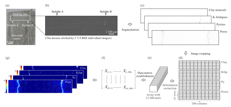
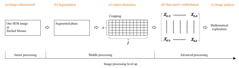
Backscatter electron analysis from scanning electron microscopes (BSE-SEM) produces high-resolution image data of both rock samples and thin-sections, showing detailed structural and geochemical (mineralogical) information. This allows an in-depth exploration of the rock microstructures and the coupled chemical characteristics in the BSE-SEM image to be made using image processing techniques. Although image processing is a powerful tool for revealing the more subtle data "hidden" in a picture, it is not a commonly employed method in geoscientific microstructural analysis. Here, we briefly introduce the general principles of image processing, and further discuss its application in studying rock microstructures using BSE-SEM image data.

| Arganda-Carreras, I., Kaynig, V., Rueden, C., et al., 2017. Trainable Weka Segmentation: A Machine Learning Tool for Microscopy Pixel Classification. Bioinformatics, 33(15): 2424–2426. https://doi.org/10.1093/bioinformatics/btx180 |
|
Boyat, A. K., Joshi, B. K., 2015. A Review Paper: Noise Models in Digital Image Processing. arXiv: 1505.03489. |
| Cheng, Q. M., 2021. IUGS' Initiative on Data-Driven Geoscience Discovery. Journal of Earth Science, 32(2): 468–470. https://doi.org/10.1007/s12583-021-1455-6 |
| Cnudde, V., Boone, M. N., 2013. High-Resolution X-Ray Computed Tomography in Geosciences: A Review of the Current Technology and Applications. Earth-Science Reviews, 123: 1–17. https://doi.org/10.1016/j.earscirev.2013.04.003 |
| De Boever, W., Derluyn, H., Van Loo, D., et al., 2015. Data-Fusion of High Resolution X-Ray CT, SEM and EDS for 3D and Pseudo-3D Chemical and Structural Characterization of Sandstone. Micron, 74: 15–21. https://doi.org/10.1016/j.micron.2015.04.003 |
| El-Gabry, E. A., Parwani, A. V., Pantanowitz, L., 2014. Whole-Slide Imaging: Widening the Scope of Cytopathology. Diagnostic Histopathology, 20(12): 456–461. https://doi.org/10.1016/j.mpdhp.2014.10.006 |
| Goldstein, J. I., Newbury, D. E., Michael, J. R., et al., 2018. ImageJ and Fiji. Scanning Electron Microscopy and X-Ray Microanalysis. Springer, New York. 187–193. https://doi.org/10.1007/978-1-4939-6676-9_13 |
| Gonzalez, R. C., Woods, R. E., 2018. Digital Image Processing (4th Ed). Pearson Education Limited, New York. 1009 |
| Hong, L., Wan, Y. F., Jain, A., 1998. Fingerprint Image Enhancement: Algorithm and Performance Evaluation. IEEE Transactions on Pattern Analysis and Machine Intelligence, 20(8): 777–789. https://doi.org/10.1109/34.709565 |
| Hou, Z. L., Fusseis, F., Schöpfer, M., et al., 2023a. Synkinematic Evolution of Stylolite Porosity. Journal of Structural Geology, 173: 104916. https://doi.org/10.1016/j.jsg.2023.104916 |
| Hou, Z. L., Woś, D., Tschegg, C., et al., 2023b. Three-Dimensional Mineral Dendrites Reveal a Nonclassical Crystallization Pathway. Geology, 51(7): 626–630. https://doi.org/10.1130/g51127.1 |
|
Jain, V., Seung, H. S., 2008. Natural Image Denoising with Convolutional Networks. Proceedings of the 21st International Conference on Neural Information Processing Systems, December 8–10, 2008, Vancouver, British Columbia, Canada. 769–776. |
|
Karras, T., Laine, S., Aittala, M., et al., 2020. Analyzing and Improving the Image Quality of StyleGAN. Proceedings of the 2020 IEEE/CVF Conference on Computer Vision and Pattern Recognition. Seattle. IEEE. 8107–8116. |
| Minaee, S., Boykov, Y., Porikli, F., et al., 2022. Image Segmentation Using Deep Learning: A Survey. IEEE Transactions on Pattern Analysis and Machine Intelligence, 44(7): 3523–3542. https://doi.org/10.1109/TPAMI.2021.3059968 |
| Prêt, D., Sammartino, S., Beaufort, D., et al., 2010. A New Method for Quantitative Petrography Based on Image Processing of Chemical Element Maps: Part Ⅰ. Mineral Mapping Applied to Compacted Bentonites. American Mineralogist, 95(10): 1379–1388. https://doi.org/10.2138/am.2010.3431 |
| Reed, S. J. B., 2005. Electron Microprobe Analysis and Scanning Electron Microscopy in Geology. Cambridge University Press, Cambridge. 215. https://doi.org/10.1017/cbo9780511610561 |
| Schindelin, J., Arganda-Carreras, I., Frise, E., et al., 2012. Fiji: An Open-Source Platform for Biological-Image Analysis. Nature Methods, 9(7): 676–682. https://doi.org/10.1038/nmeth.2019 |
| Sonka, M., Hlaváč, V., Boyle, R., 2013. Image Processing Analysis and Machine Vision. Springer, New York. 554 |
| Swamy, S., Kulkarni, P. K., 2020. A Basic Overview on Image Denoising Techniques. Int. Res. J. Eng. Technol., 7(5): 850–857 |
| Tschegg, C., Hou, Z. L., Rice, A. H. N., et al., 2020. Fault Zone Structures and Strain Localization in Clinoptilolite-Tuff (Nižný Hrabovec, Slovak Republic). Journal of Structural Geology, 138: 104090. https://doi.org/10.1016/j.jsg.2020.104090 |
| Wang, Z., Bovik, A. C., 2006. Modern Image Quality Assessment. Morgan & Claypool Publishers, California. 146 |
| Xu, J., Yang, L., Wu, D. P., 2010. Ripplet: A New Transform for Image Processing. Journal of Visual Communication and Image Representation, 21(7): 627–639. https://doi.org/10.1016/j.jvcir.2010.04.002 |
| Zehner, B., Börner, J. H., Görz, I., et al., 2015. Workflows for Generating Tetrahedral Meshes for Finite Element Simulations on Complex Geological Structures. Computers & Geosciences, 79: 105–117. https://doi.org/10.1016/j.cageo.2015.02.009 |
| Zhang, L., Qiu, K. F., Hou, Z. L., et al., 2021. Fluid-Rock Reactions of the Triassic Taiyangshan Porphyry Cu-Mo Deposit (West Qinling, China) Constrained by QEMSCAN and Iron Isotope. Ore Geology Reviews, 132: 104068. https://doi.org/10.1016/j.oregeorev.2021.104068 |
Figures(3)
Copyright © 2013-2020 Journal of Earth Science 鄂ICP备15021562号-2
Tel: +86-27-67885075 Fax: +86-27-67885075 E-mail: xbb@cug.edu.cn
Address: Editorial Office of Journal, China University of Geosciences, Yujiashan, Wuhan, Hubei 430074, P. R. China
Supported by:
Beijing Renhe Information Technology Co. Ltd
E-mail:
info@rhhz.net


 DownLoad: